Anatomy of the human heart: chambers, valves and vessels
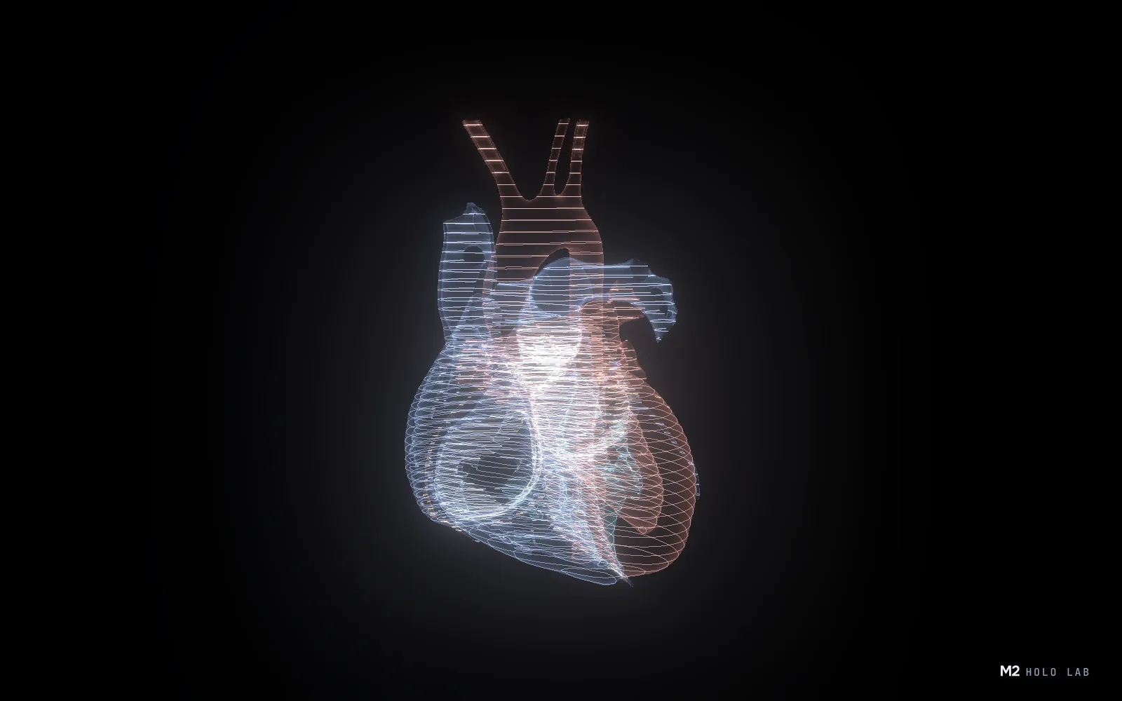
The heart is a muscular pump slightly larger than a fist, weighing about 200–425 g. Inside it has four chambers and four valves. The right side pumps blood to the lungs, the left side to the whole body. Below is the structure of the heart part by part, on a 16-part 3D model you can take apart with your hands in M2.
Where the heart is
The heart sits in the chest between the lungs, behind the breastbone and slightly left of centre. It is wrapped in a protective sac, the pericardium. Its wall has three layers: the inner endocardium, the thick muscular myocardium and the outer epicardium.
The four chambers
The upper chambers, the atria, receive blood. The lower chambers, the ventricles, push it out of the heart. A wall called the septum divides the right and left sides, so blood from the two sides does not mix.
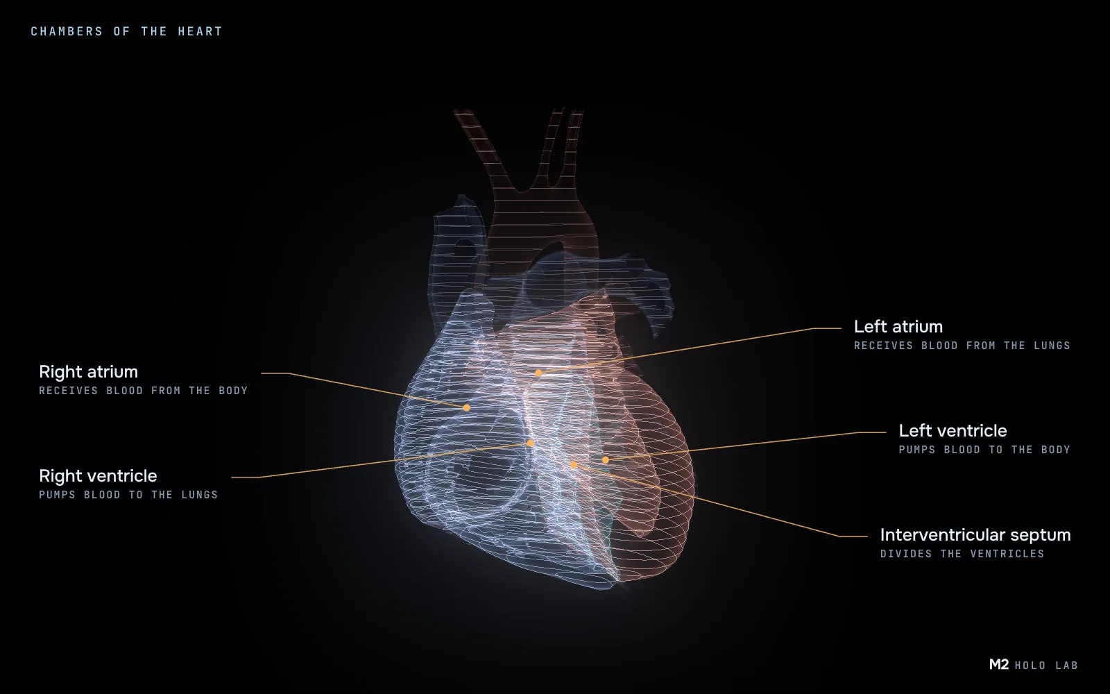
- Right atrium receives oxygen-poor blood from the body through the superior and inferior venae cavae. Its wall holds the sinoatrial node, the heart's natural pacemaker, where every beat starts.
- Right ventricle pumps blood to the lungs. Pressure in the lung vessels is low, so its wall is thinner than the left one, usually 3–5 mm.
- Left atrium receives oxygen-rich blood from the lungs through the pulmonary veins, usually four of them.
- Left ventricle is the main pump: it pushes blood into the aorta and on to the whole body. Its wall is the thickest in the heart, about 1 cm, and the pressure it builds is about 120 mmHg.
- Interventricular septum is the muscular wall between the ventricles. It keeps the blood of the two sides apart and takes part in every contraction.
The four valves
Valves let blood flow in one direction only. Two sit between the atria and the ventricles, two at the exits of the ventricles.
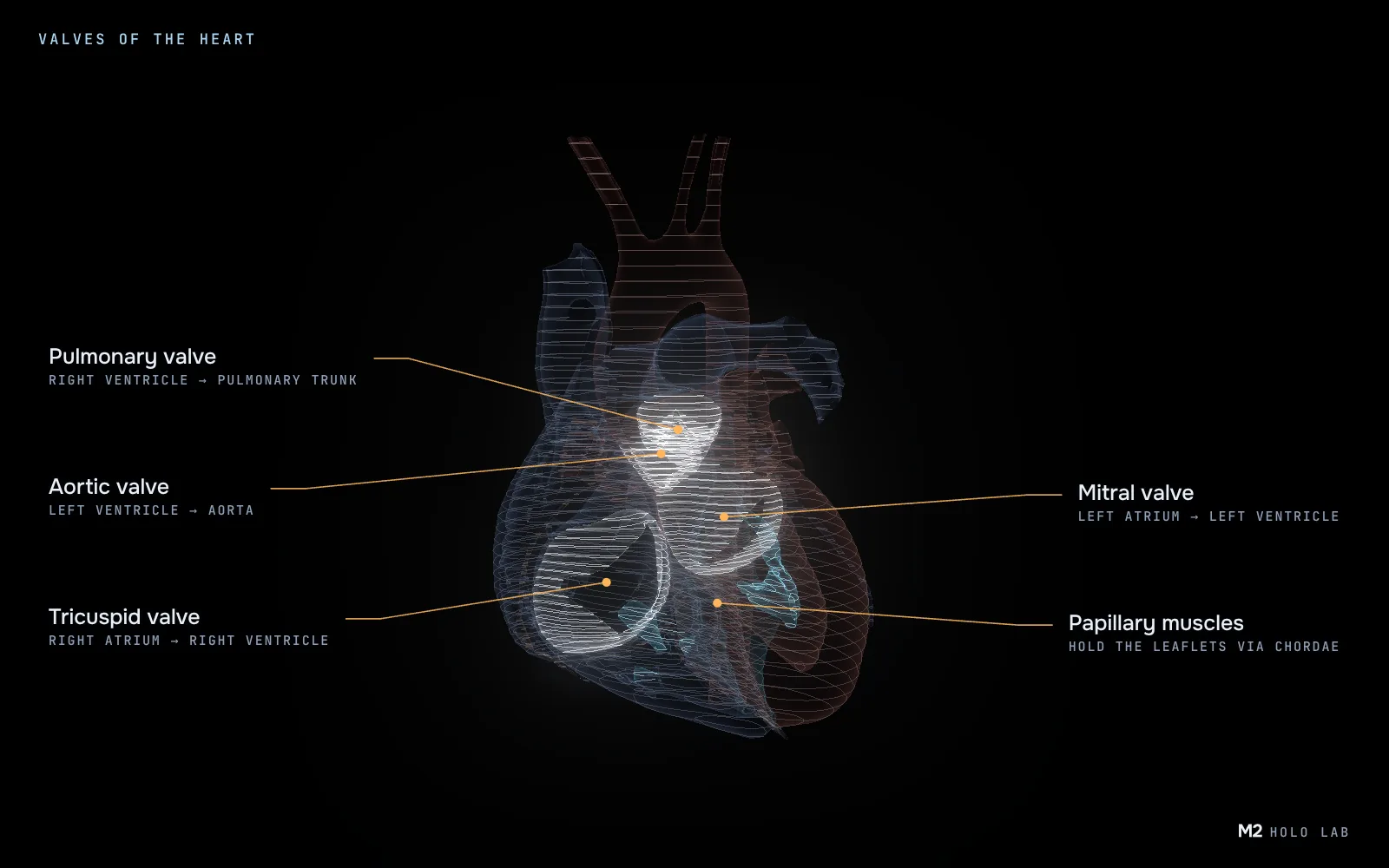
| Valve | Where it sits | Leaflets |
|---|---|---|
| Tricuspid | right atrium → right ventricle | 3 |
| Pulmonary | right ventricle → pulmonary trunk | 3 semilunar |
| Mitral | left atrium → left ventricle | 2 |
| Aortic | left ventricle → aorta | 3 semilunar |
The leaflets of the tricuspid and mitral valves are held by chordae tendineae, strong cords attached to the papillary muscles inside the ventricles. When a ventricle contracts, the papillary muscles pull the cords tight, so the leaflets are not pushed back into the atrium.
The familiar "lub-dub" is the sound of valves closing: first the mitral and tricuspid valves, then the aortic and pulmonary valves.
The great vessels
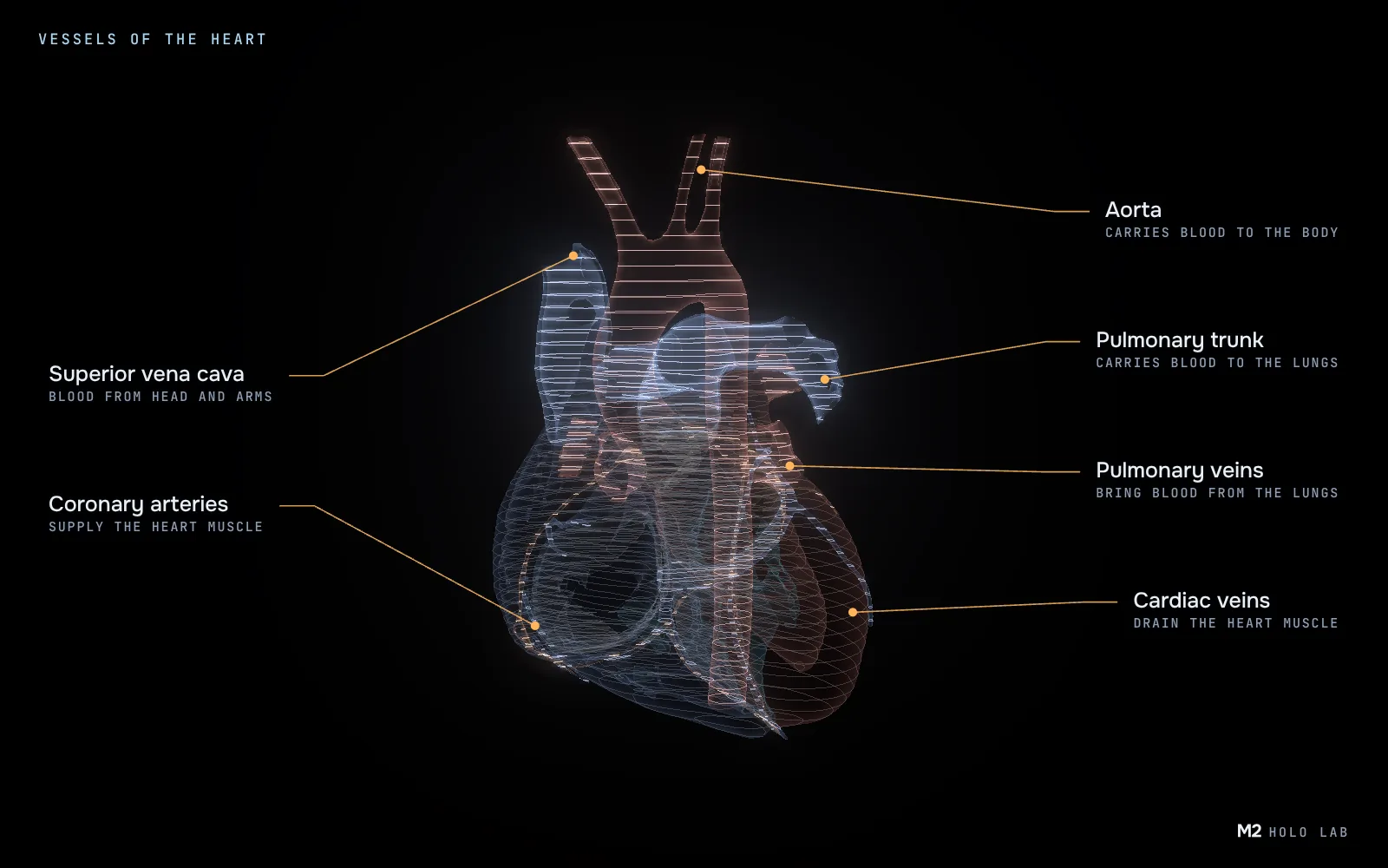
- Superior and inferior venae cavae bring blood from the upper and lower body into the right atrium.
- Pulmonary trunk leaves the right ventricle and splits into the right and left pulmonary arteries. In adults these are the only arteries that carry oxygen-poor blood.
- Pulmonary veins carry oxygen-rich blood from the lungs to the left atrium, the only veins that do.
- Aorta leaves the left ventricle. It is the largest artery in the body, about 3 cm across. Vessels to the head and arms branch off its arch, then it runs down along the spine.
The heart's own vessels
The heart feeds itself through the coronary arteries. They branch off the root of the aorta just above the aortic valve and run over the surface of the heart. The left coronary artery splits into the left anterior descending and circumflex arteries; the right one supplies the right side of the heart and the back of the septum. A blocked coronary artery is the main cause of a heart attack.
Cardiac veins collect used blood from the heart muscle. They join into the coronary sinus, which empties into the right atrium.
The path of blood through the heart
- Blood from the body arrives through the venae cavae in the right atrium.
- It passes the tricuspid valve into the right ventricle.
- The right ventricle pushes it through the pulmonary valve into the pulmonary arteries.
- In the lungs, blood gives up carbon dioxide and picks up oxygen.
- Through the pulmonary veins it returns to the left atrium.
- It passes the mitral valve into the left ventricle.
- The left ventricle pushes it through the aortic valve into the aorta, and from there to the whole body.
Both sides work at the same time: first both atria contract, then both ventricles. So the right side sends blood to the lungs at the very moment the left side sends it to the body.
How we built this model
The model comes from HuBMAP, an open anatomical atlas. The source meshes are very detailed, so for the browser we simplified them, split the heart into 16 parts and compressed them, so the model loads quickly on an ordinary laptop. The horizontal lines in the illustrations are slice contours: they keep the hologram readable even where parts overlap.
The colours follow the blood, not a textbook atlas: blue sections carry oxygen-poor blood, red ones oxygen-rich blood. So the model shows at a glance which half of the heart works for the lungs and which for the whole body.
Not everything worked the first time:
- Thin vessels. The coronary arteries and pulmonary veins were harder to grab by hand than other parts: 14 tries out of 16 in our tests. That's why any part can also be picked from the list.
- Labels. At first all 16 parts were labelled at once, and the labels covered the heart itself. Now a label appears only on the part your hand points at.
- Dark spots on Macs. On Apple graphics chips, dark holes appeared on the surface of the parts: in rare points the lighting formula returned "not a number". It never showed on our test machine; we found it from a screen recording made on a real Mac.
Studying the heart in 3D
In M2 the heart model comes apart into 16 parts, grouped the same way as in this article: chambers, valves, great vessels and the heart's own vessels. You can pull a part out with a pinch, turn it in your hand and open it full screen. In close-up the model turns semi-transparent, so you can look inside a chamber or a valve. Every part has a short explanation card.
The model is built on data from the Human Reference Atlas (HuBMAP), made from the Visible Human Project of the US National Library of Medicine. It runs in the browser with an ordinary webcam, no VR headset.
Questions and answers
How many chambers does the human heart have?
Four: the right and left atria at the top and the right and left ventricles at the bottom. The atria receive blood, the ventricles push it out of the heart.
How many valves are in the heart?
Four: tricuspid, pulmonary, mitral and aortic. They let blood flow in one direction only.
Why is the left ventricle wall thicker than the right?
The left ventricle pumps blood to the whole body at about 120 mmHg, while the right one pumps only to the lungs, where pressure is much lower. So the left wall is about 1 cm thick and the right one 3–5 mm.
What supplies blood to the heart itself?
The coronary arteries. They branch off the root of the aorta and run over the heart's surface; used blood is collected by the cardiac veins and the coronary sinus.
How much does a human heart weigh?
Usually about 200–425 g, slightly larger than a fist.
Sources
- NHLBI (NIH): How the Heart Works
- Texas Heart Institute: Heart Anatomy
- Cleveland Clinic: Heart
- Cleveland Clinic: Heart Valves
- Radiopaedia: Chordae tendineae
- HuBMAP Human Reference Atlas: 3D Reference Organ Set, doi:10.48539/HBM447.LMKF.747
Illustrations are rendered from the M2 heart model. Facts are checked against the sources listed below. This article is for learning and is not medical advice.